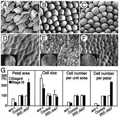
Figure 3.
Loss- or gain-of-ANT-function influences the extent of cell proliferation during organogenesis. (A–C) Fully differentiated cells (×135) with centripetal ridges characteristic of the adaxial epidermis of mature petals at stage 15. (D–F) Undifferentiated epidermal cells (×450) from the adaxial, distal portion of mid-stage 9 petals (Insets, ×45). (A and D) ant-1 petal. (B and E) Control (wild type). (C and F) 35S::ANT. (G) Comparison in petal area, cell size, cell number per unit area, and numbers per petal. Percentages of results from ant-1 and 35S::ANT petals to those from control petals are shown. Bars indicate SD. The analysis was performed with the abaxial, distal portion of petals by using more than 20 flowers.

