Figure 2.
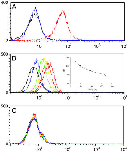
SCS-873 directs mAb 38C2 to cells expressing integrin αvβ3 and αvβ5. (A) Flow cytometry histogram showing the binding of mAb 38C2 to human melanoma M21 cells in the presence of a twice equimolar concentration of SCS-873 (red). MAb 38C2 and SCS-873 were mixed before cell binding. FITC-conjugated goat anti-mouse polyclonal antibodies were used for detection. Like mAb 38C2 alone (blue), SCS-873 alone (data not shown) was indifferent from the background signal of secondary antibodies alone (black). The y axis gives the number of events in linear scale, the x axis the fluorescence intensity in logarithmic scale. (B and C) Flow cytometry histogram showing the binding of mouse sera to human melanoma M21 cells. Shown are 1:100 dilutions of sera from mice injected i.p. with 1 mg SCS-873 and i.v. with either 1 mg mAb 38C2 (B) or 1 mg isotypic control mAb (C). Sera were prepared from eye bleeds taken 24 h (red), 48 h (orange), 72 h (yellow), 96 h (green), and 168 h (blue) after the injections. FITC-conjugated goat anti-mouse polyclonal antibodies were used for detection. Typical results based on three individual mice in each treatment group are shown. Inset in B shows the decline of the mean fluorescence intensity (MFI) over time.

