Figure 6.
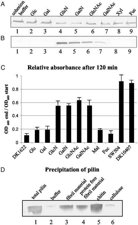
Effect of monosaccharides on TFP retraction. Cell-surface pilin was assayed as described for Fig. 3. (A) SW504 (4 × 10⁸ cells) was incubated at 32°C for 150 min with cohesion buffer (lane1); 100 mM glucose (Glc), galactose (Gal), GlcN, GalN, GlcNAc, GalNAc, xylose (Xyl) (lanes 2–8), or fucose (Fuc) (as negative control, lane 9) and surface pilin was immunoblotted. All sugar solutions had been adjusted to a pH of 7 and cell viability was tested after the incubation, showing no noticeable difference when compared with the cohesion buffer control. (B) DK1622 (4 × 10⁸ cells) was incubated at 32°C for 150 min with 100 mM designated sugars and cell-surface pilin was immunoblotted. Lanes 1–9, same as in A. Data presented in A and B are representative for triplicate experiments. (C) Cohesion inhibition of wild-type DK1622 on the addition of different sugars at a final concentration of 50 mM. Mal, maltose; other abbreviations are the same as in A. SW504 and DK10407 were used as negative controls. OD600 was taken every 10 min for 120 min. The OD600 of each sample after 120 min (OD600 end), relative to its OD600 at the beginning of the assay (OD600 start), is shown in the graph. Data represent the mean ± SD of three experiments. (D) Cell-surface pilin was sheared off from 5 × 10⁸ SW504 cells and the total pilin amount as prepared by MgCl2 precipitations shown in lane 1. Same amounts of isolated pilin were also precipitated with cohesion buffer (lane 2), fibril material (0.2 mg/ml carbohydrate, lane 3), Pronase-treated fibril material (0.2 mg/ml carbohydrate, lane 4), chitin suspension (0.2 mg/ml, lane 5, and cellulose suspension (0.2 mg/ml, lane 6).

