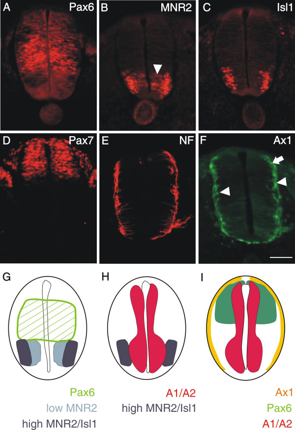
Figure 3.
PlexinA1 and A2 are already expressed in neural precursors. At stage 18, the homeodomain protein Pax6 labels precursor cells in the intermediate zone of the developing spinal cord (A; [60]). Its ventral expression boundary that is defined by the morphogen Shh released from the floor plate reaches the area with the progenitors of motor neurons expressing MNR2 (B and G). Low levels of MNR2 proteins are seen in progenitor cells located medially (arrowhead in B), where motor neurons are born. MNR2 protein persists and accumulates in postmitotic motor neurons expressing Isl1 (C). In the dorsal spinal cord, Pax7 expression marks a population of precursor cells that give rise to dorsal interneuron subpopulations (D; [37]). Note the decline of Pax7 staining toward the periphery of the spinal cord. Even at stage 19, postmitotic neurons expressing neurofilament proteins are found only at the peripheral margin of the spinal cord (E). At this stage, the dorsolateral population of commissural axons, characterized by the expression of Axonin-1 (arrow in F), starts to extend axons toward the floor plate [38]. At the same, more ventrally located interneurons expressing Axonin-1 also extend axons but their pathway has not been characterized in detail (arrowheads). A comparison of the expression of Pax6 (green hatched area), MNR2 (light and dark blue, characterizing low and high protein levels, respectively) and Isl1 (dark blue) is shown in G. For clarity the mRNA expression domains of plexinA1 and A2 have been added in H (red). Significant overlap was found between the plexin expression domain and early motor neurons characterized by MNR2 staining (H). In the dorsal spinal cord domains of Pax7 (green) and Axonin-1 protein expression (yellow) are compared to the domains expressing plexinA1 and A2 mRNA in I. Plexins do not extend to the periphery of the neural tube where postmitotic neurons expressing neurofilament or axonin-1 are found. Bar in A through F 50 μm.

