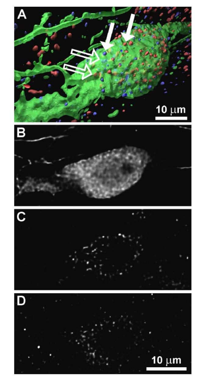
Fig. 3.

Immunocytochemical triple staining for OT (green), vasoactive intestinal polypeptide (VIP) receptor (red), and PRL receptor (blue) of a sample cell in paraventricular nucleus (PVN). A: 3-dimensional reconstruction of a PVN cell from a stack of optical sections taken by confocal laser-scanning microscope. In addition to receptors located on cell surface (filled arrows), there was also cytoplasmic immunopositve receptor staining (open arrows). B–D: display of a 2-dimensional scan from stack of optical sections on level of cell soma, divided into 3 recorded channels; B = OT (A, green), C = VIP receptor (A, red), D = PRL receptor (A, blue).
