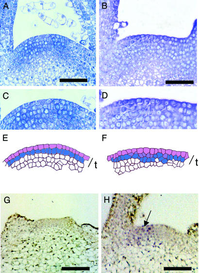
Figure 2.
Induction of phragmoplastin gene expression leads to altered cell division pattern in the apical meristem. (A) Longitudinal section through the meristem of a control apex. (B) Longitudinal section through the meristem of a Tet∷SDL12-1 plant in which microinduction has been performed. (C) Detail from the meristem shown in A. (D) Detail from the meristem shown in B. (E) Cellular outline of the meristem shown in C. The outer cells of the tunica (t) have been colored to highlight the layered cellular architecture. Cells connected by anticlinal cell walls are indicated by separate colors. (F) Cellular outline of the meristem shown in D. The outer cells of the tunica (t) have been colored to highlight the occurrence of nonanticlinal cell divisions at various positions within the tunica. Cells connected by anticlinal cell walls are indicated by separate colors. (G) In situ hybridization using a sense probe for SDL12 of a Tet∷SDL12-1 apex induced at the I2 position with Ahtet. No signal is visible. (H) In situ localization as in G except an antisense probe for SDL12 was used. Signal (blue/violet) is seen on one flank of the meristem (arrow). (Bars in A and B = 50 μm; bars in G and H = 100 μm.)

