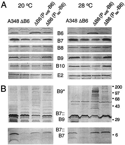
FIG. 4.
Accumulation of the VirB6 through VirB10 proteins as a function of altered VirB6 production and growth temperature. (A) Immunoblot analysis of total protein samples electrophoresed under reducing conditions; blots were developed with antisera specific for the proteins listed in the center. (B) Immunoblot analysis of protein samples electrophoresed under nonreducing conditions; top panels were developed with anti-VirB9 antiserum to show the VirB7::VirB9 heterodimer (B7::B9) and higher-order VirB9 complexes (B9*), and bottom panels were developed with anti-VirB7 antiserum to show the VirB7 homodimer (B7::B7). Strains: A348, wild-type; ΔB6, PC1006; ΔB6(PvirB-B6), PC1006(pSJB964); ΔB6(PlacZ-B6), PC1006(pXZB61). Total cellular proteins from equivalent numbers of cells were subjected to SDS-PAGE and immunoblot analysis. Molecular size markers (in kilodaltons) are shown at right.

