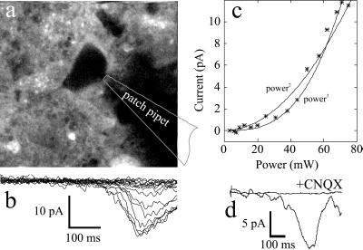
Figure 5.
Responses to two-photon uncaging of Bhc-glu. (a) Two-photon fluorescence image of a hippocampal neuron bathed in 0.5 mM Bhc-glu. (b) Whole-cell currents recorded in response to full-field raster scans like the one used for the fluorescence image (2 ms per line, 256 lines) with the beam power between 0 to 75 mW. (c) Dependence of peak current on beam power. The curves indicate fits by eye to second- and third-power functions. (d) Block of uncaging-evoked currents by a glutamate receptor antagonist (laser power 80 mW). Traces show responses at the same location in the absence and presence of 50 μM CNQX (6-cyano-7-nitroquinoxaline-2, 3-dione), an antagonist of non-N-methyl-d-aspartate receptors.

