Fig. 1.
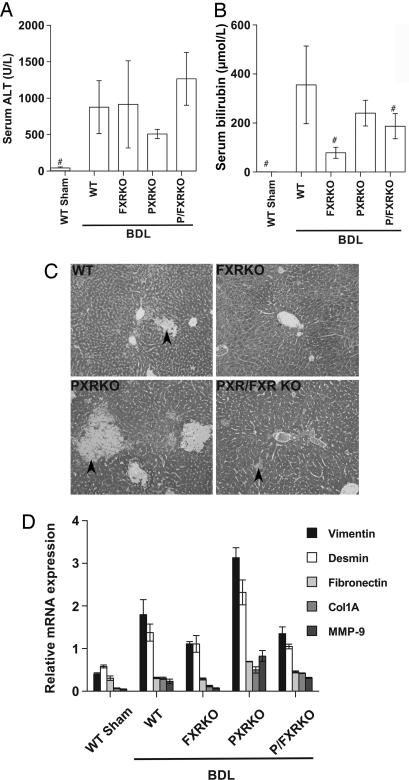
Deletion of PXR or FXR results in differential patterns of liver injury after BDL. Serum and liver samples were collected 6 days after BDL or sham operation in WT, FXRKO, PXRKO, and P/FXRKO mice. (A) Serum ALT was significantly elevated in all BDL animals compared with shams. (B) Total serum bilirubin was significantly elevated in WT, PXRKO, and P/FXRKO BDL mice relative to sham and was significantly lower in FXR-null mice than WT after BDL. #, P < 0.01 relative to WT BDL. (C) Livers were fixed for histological examination and stained with Gomori’s Trichrome to evaluate liver injury and necrosis. WT livers show modest areas of bile infarct/necrosis (arrowheads); PXRKO mice have increased areas of bile infarct/necrosis compared to WT; FXRKO and P/FXRKO mice have a reduction in bile infarcts, but disseminated liver cell necrosis and mild to moderate hepatocyte steatosis is still observed. (D) Effects of BDL on hepatic expression of genes involved in fibrosis and tissue remodeling. Relative mRNA expression of vimentin, desmin, fibronectin, collagen 1A (Col1A), and matrix metalloproteinase 9 (MMP-9) were examined by real-time RT-PCR and normalized for U36B4 expression.

