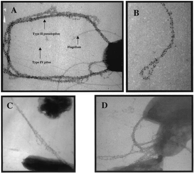
FIG. 3.
XcpT pseudopilin is a major component of the type II pseudopilus. Shown are the results of TEM analysis of PAO1/pMTWT after immunogold labeling with antibodies raised against XcpT. (A, C, and D) The type II pseudopili are seen attached to bacterial cells. (B) A type II pseudopilus with a loop structure is seen enlarged. In panel A, the positions of a type IV pilus, the flagellum, and a type II pseudopilus have been indicated.

