Fig. 1.
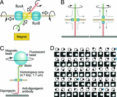
Observation of Holliday junction DNA rotation by RuvA–RuvB. (A) Model of branch migration by RuvA–RuvB. Branch migration in solution is depicted. The rotational direction of each DNA strand is indicated by an arrow. Thick arrows indicate the direction of DNA translocation. (B) One end of cruciform DNA is fixed to the glass surface. The RuvB hexameric rings bind to both sides of the junction along the horizontal DNA, the rotation of the lower DNA is restrained, and the right-handed helical rotation of DNA is observed from above (Left). In contrast, when the RuvB hexameric ring binds to both sides of the junction along the vertical axis, left-handed helical rotation of DNA is observed (Right). (C) Observation system (not to scale). The magnetic bead was pulled upwards by a disk-shaped neodymium magnet. The direction of the magnetic field was vertical and did not prevent bead rotation. Daughter fluorescent beads served as markers of rotation. (D) Snapshots of rotating beads at 100-ms intervals at an ATP concentration of 50 μM. The moving white spot is a daughter fluorescent bead. Diagrams show their relative positions. Blue arrowheads indicate completion of a turn.

