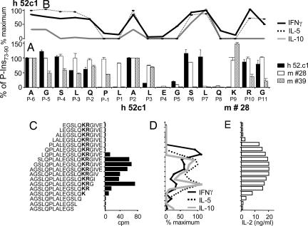
Fig. 2.
Determination of DR4/TCR contacting positions of P-Ins73-90 and influence of the flanking amino acids on the activation and cytokine secretion of the human T cell clone. (A) Reactivity of the human T cell clone 52c1 (black bars) and two mouse hybridoma, #28 (open bars) and #39 (dotted bars) to a panel of alanine-substituted peptides of P-Ins73-90. The x axis displays the sequence of the peptide and assigned positions (P) concerning core HLA/TCR-binding residues. Data used for the human T cell clone express the cpm of each of the individual peptides in relation to P-Ins73-90 (100% value). For the murine T cell hybridoma, IL-2 production (pg/ml) has been similarly related to the IL-2 production of the P-Ins73-90 (100% value). Bars represent responses to a variant peptide in which that residue alone was substituted with alanine. (B) Cytokine reactivity of the human T cell clone to the panel of alanine-substituted peptides of P-Ins73-90. Maximum IFNγ response was 459 pg/ml, for IL-5 880 pg/ml, and for IL-10 13 pg/ml. (C–E) A panel of 18 N- or C-terminally truncated peptides was used to identify a minimal core epitope of P-Ins73-90 by the human T cell clone (C) and by the mouse hybridoma #39 (E). Cytokine secretion profile of the human T cell clone after stimulation with truncated peptides of P-Ins73-90 (D). In this experiment, maximum response for IFNγ was 1,154 pg/ml; for IL-5, response was 2,817 pg/ml; and for IL-10, response was 72 pg/ml.

