Fig. 1.
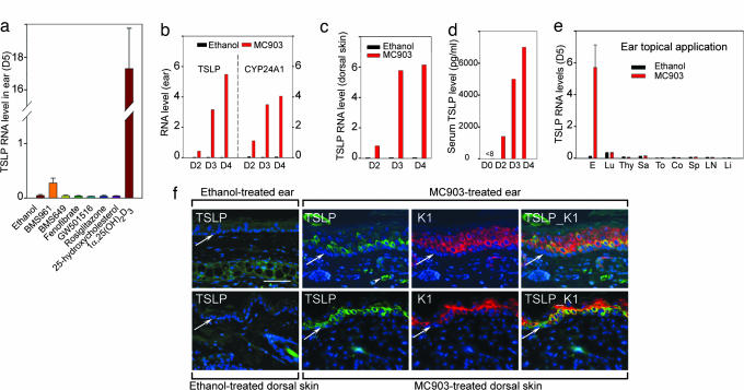
Skin topical application of 1α,25-(OH)2D3 and MC903 activates TSLP expression in epidermal keratinocytes. (a) TSLP RNA at day 5 in ears topically treated with NR agonists (4 nmol). (b–e) Induction of TSLP expression is a rapid and local skin effect. TSLP and CYP24A1 RNA levels in an ethanol-treated left ear and MC903-treated right ear (b), TSLP RNA levels in ethanol-treated and MC903-treated dorsal skin at days 2, 3, and 4 (c), and increased serum TSLP levels at days 2, 3, and 4 (d), in contrast to undetectable level (<8 pg/ml) at day 0 (before treatment). Data are representative of three independent experiments. D, day. (e) TSLP RNA levels at day 5 in ear (E), lung (Lu), thymus (Thy), salivary gland (Sa), tongue (To), colon (Co), spleen (Sp), lymph node (LN), and liver (Li) of mice topically treated by ethanol or MC903 on ears. (f) IHC of TSLP (green) at day 4 in sections of ear (Upper) and dorsal skin (Lower) topically treated with ethanol or MC903. The same sections from MC903-treated skin were stained with keratin 1 antibody (K1, red), and overlaid images of TSLP and K1 staining are shown, as indicated. Blue corresponds to DAPI staining of nuclei. The white arrowhead points to autofluorescent erythrocytes, and white arrows point to the dermal/epidermal junction. (Scale bar, 50 μm.)

