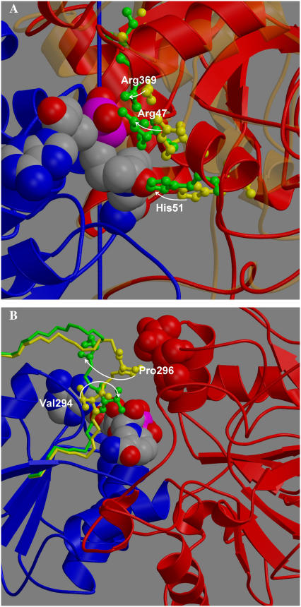
FIGURE 3.
Views on key regions in LADH. (A) The binding domain is in blue, and NAD+ in space-filling model. The movement of Arg47, His51, and Arg369 in going from the open (yellow) to closed x-ray structure (green) is indicated by arrows. The catalytic domain is in bold red (closed) and faint orange (open). (B) Loop in open (yellow) and closed (green) conformation. Rotation of Val294 (ball and stick model) to interact with NAD+ (space filling) by rotation about φ-angle of Gly293 would cause Pro296 (ball and stick model) to move away from contacts 56 and 57 (space filling, red) on catalytic domain, thereby allowing it to close.

