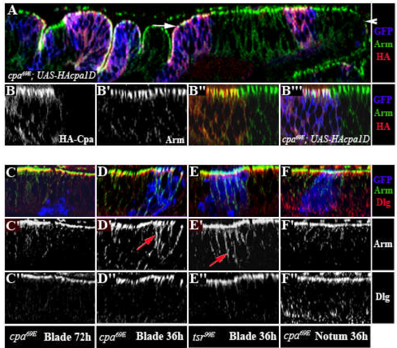
Figure 3. cpa maintains the localization of adherens junction components.

(A–F) Optical cross sections through third instar wing imaginal discs, stained with anti-Arm (red in A, B”,B”‘, C,D,E,F or white in B’,B”,C’,D’,E’,F’) and anti-HA, reflecting UAS-HA-cpa expression (green in A,B”,B”‘ or white in B) or anti-Dlg (green in C,D,E,F or white in C”,D”,E”,F”). (A–B) cpa69E mutant clones overexpressing HA-tagged full-length cpa, positively labelled with GFP (blue in A,B”‘). The white arrow in (A) defines the wing blade region. (B–B”‘) Magnification of the blade primordium. HA-cpa accumulates at the apical membrane, partly co-localizes with Arm in all regions of the wing disc and rescues extrusion of cpa mutant clones in the wing blade primordium. (C–D, F) cpa69E mutant clones positively labeled with GFP (blue in C,D,F) in the wing blade (C,D) or notum (F) primordium, induced both at second or early third instar and dissected at the late third instar stage either 60 hours (C) or 36 hours (D,F) after clone induction. While we could recover mutant clones within the disc epithelium 36 hours after clone induction, all mutant cells were extruded by 60 hours. (E) tsr99E mutant clones in the blade primordium, dissected 36 hours after clone induction and positively labeled with GFP (blue in E). Arm is mislocalized to basolateral regions in extruding cpa mutant cells and in tsr mutant cells that are maintained within the epithelium (red arrows in D’ and E’). Following extrusion of cpa mutant cells, expression of both Arm and Dlg are lost (C).
