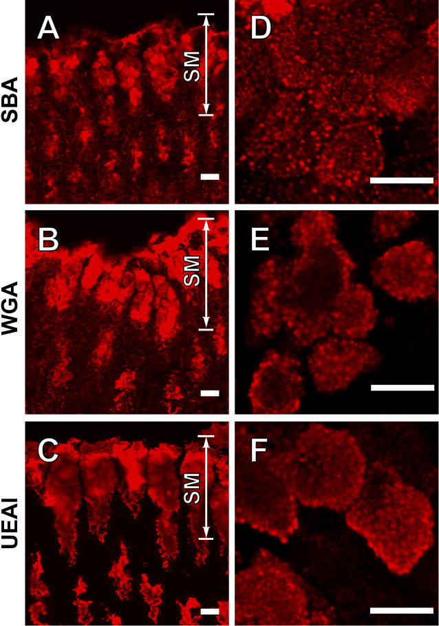
Figure 1. Three Lectins Strongly Stain the Intracellular Content of Gastric Surface Mucous Cells.
Frozen sections of the fundic portion of the mouse stomach were stained with rhodamine-labeled SBA (A), WGA (B), or UEA1 (C). Note staining of the surface mucous (SM) cells, which are the mucus-producing population of the gastric epithelium. Primary cultures of gastric surface mucous cells (asterisks) were also stained with SBA (D), WGA (E), or UEA1 (F). Confocal imaging reveals labeling of a spherical, large (~ 1-μm diameter), abundant organelle, the mucus granule. Bars represent 10 μm.

