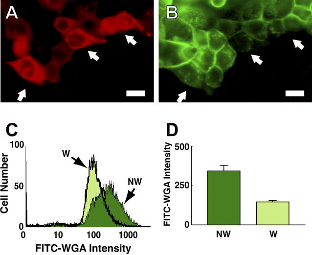
Figure 4. Cells That Survive a Plasma Membrane Disruption Are Depleted of Intracellular Mucus.
(A) MKN28 cell monolayer substrata were scratched with the tip of a needle, denuding cells and wounding many of those bordering on the denudation site. Those cells that resealed the plasma membrane disruptions that were thus created trap in their cytoplasm the Texas red-labeled (TRDx) dextran present during the scratch injury, and their cytosol is consequently fluorescent (arrows).
(B) Images of the same cells after staining of intracellular mucus with FITC-WGA. Note that those cells heavily labeled with the TRDx-dextran, which suffered and repaired large plasma membrane disruptions, apparently stain more lightly with the FITC-WGA.
(C) Flow cytofluorometic analysis of FITC-WGA staining in MKN28 populations that were wounded by syringing (W) or were undisturbed (NW) prior to intracellular staining of mucus with FITC-WGA. Note the downward shift in population fluorescence of the wounded relative to the undisturbed population.
(D) The mean value of wounded (W) and undisturbed (NW) population fluorescence as measured by flow cytofluorometry (n = 3; p < 0.05). Bars represent 20 μm.

