Figure 1.
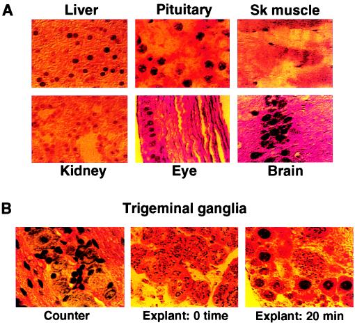
Distinct localization of the C1 factor in sensory neurons. (A) Sections of representative mouse tissues were stained with purified anti-C1 sera. In each case, the protein is detected in the cell nucleus. (B) Mouse trigeminal ganglia were excised and immediately fixed (0 time) or were explanted into media for 20 min before fixation (20 min). Representative sections from the ganglia were counterstained with hematoxylin to visualize the clustered neurons and neuronal nuclei (Counter) or were stained with purified anti-C1 sera. Although the protein is detected in cytoplasmic punctate structures at 0 time, it is primarily localized to the nucleus of explanted ganglia neurons. Control antibodies directed against neurofilaments demonstrated that the tissue was intact and permeable to antibody staining (data not shown).

