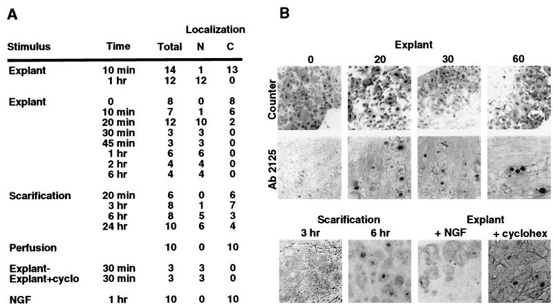
Figure 2.
Correlation of C1 localization and HSV reactivation. (A) Trigeminal ganglia were explanted and incubated in media for various times before fixation (Explant), excised at various times after eye scarification (Scarification), excised after fixation-perfusion (Perfusion), incubated in the presence of NGF (7.5S NGF, 10–100 ng/ml), or incubated in the presence of cyclohexamide (+cyclo, 100 μg/ml). The treated ganglia were fixed, embedded, and stained with anti-C1 sera or control sera as described. The total number of ganglia in each experiment is shown with the number that exhibit nuclear (N) or cytoplasmic (C) phenotypic localization of the C1 factor. Each ganglion contains 200–400 neurons per section. Designation as nuclear indicates that 10–30% of the neurons in a ganglion exhibited nuclear staining, whereas cytoplasmic exhibit 0.2–0.8% nuclear staining. Three to five sections from each embedded ganglion were stained to determine an experimental point. (B) Representative sections from the experimental sets listed above. Sections were counterstained to visualize intact neuronal nuclei (Counter) or were stained with anti-C1 Ab2125 serum.

