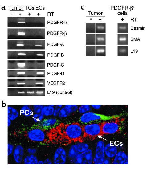
Figure 3.
Identification of the cell types expressing PDGF ligands and receptors in pancreatic islet carcinomas. (a) Primary tumors were fractioned into constituent cell types by flow cytometry. RNA was isolated from unsorted and sorted populations and analyzed by RT-PCR. Pancreatic tumors of end-stage Rip1Tag2 mice were excised and enzymatically dispersed with collagenase into single cells. The cell suspension was incubated with Ab’s for CD31 and Gr1 and Mac1. Endothelial cells were collected by FACS as a CD31+, Gr1–, Mac1– population, whereas tumor cells were gated by size and collected as unlabeled with these three Ab’s. Inflammatory cells were collected as Gr1+, Mac1+; these cells did not express PDGF ligands or receptors (not shown). (b) Tumor sections (prepared as in Figure 2) were costained with anti–PDGFR-β-FITC (1:200) and anti–CD31-rhodamine to reveal PDGFR-β-expressing cells in green and endothelial cells in red. (c) PDGFR-β+ cells from tumors were isolated by FACS (PDGFR-β Ab, 1:50), RNA isolated, and analyzed by RT-PCR for pericyte markers. ECs, endothelial cells; TCs, tumor cells; PCs, perivascular cells.

