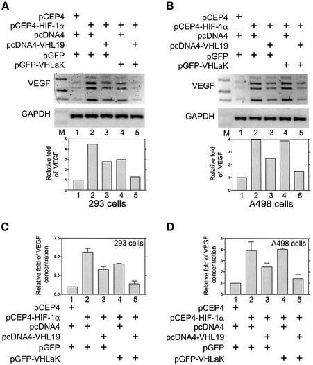
Fig. 5. The VHLaK protein inhibits VEGF mRNA and protein expression in the presence of pVHL. (A and B) the VHLaK protein inhibits HIF-1α- induced VEGF mRNA expression in the presence of pVHL. HEK 293 cells (A) and A498 cells (B) were transfected with the plasmids as indicated. Thirty-six hours after transfection, total RNA was extracted and semi-quantitative RT–PCR was performed using primers specific for VEGF or GAPDH. Note that four bands were observed representing four VEGF isoforms (VEGF 121, 165, 189 and 206). The densities of VEGF bands were quantified and normalized with the density of GAPDH bands. Molecular size markers were loaded in lane M. (C and D) The VHLaK protein inhibits HIF-1α-induced VEGF protein expression in the presence of pVHL. HEK 293 cells (C) and A498 cells (D) were transfected with the plasmids as indicated. Thirty-six hours after transfection, aliquots of medium were collected for VEGF ELISA and results were normalized for cell numbers. Histograms represent the results of three independent experiments.

An official website of the United States government
Here's how you know
Official websites use .gov
A
.gov website belongs to an official
government organization in the United States.
Secure .gov websites use HTTPS
A lock (
) or https:// means you've safely
connected to the .gov website. Share sensitive
information only on official, secure websites.
