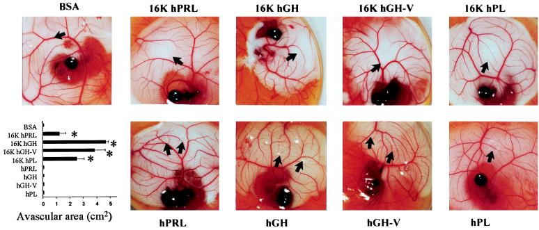
Figure 2.
Early-stage CAM assay showing inhibition of angiogenesis in the CAM: Representative examples of CAM are taken from a typical experiment. The disks are visible by light reflection, and the black arrow shows the border of the disk or the border of the avascular area, if present. The lower left panel shows the quantification of the assay performed by measuring the area devoid of capillaries in the region surrounding the disk. Values are means ± SEM. ∗, P < 0.01 vs. BSA.

