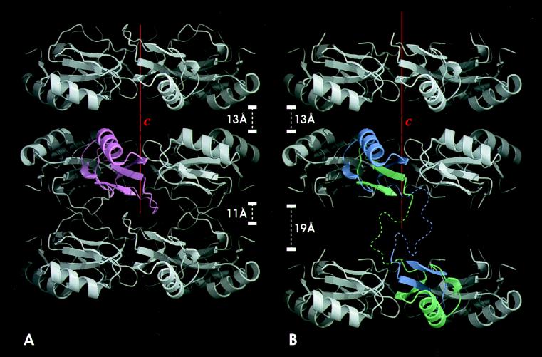
Figure 3.
The quaternary structure and crystal packing of wild-type CI2 (A) compared with that of dimeric CI2-Q4i (B). Three hexameric ring layers are shown. In B, a CI2-Q4i dimer is colored, and, in A, a CI2 monomer is colored. In the CI2-Q4i structure, one of the spaces between the layers is expanded to 19 Å, compared to 11 Å in the wild-type. The color coding is the same as in Fig. 2. The dotted lines represent the inferred positions of the disordered residues. The red boxes are the unit cells. This view is along the b axis.

