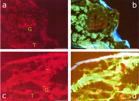
FIG. 2.
FISH with microscopic transverse (a and b) and sagittal (c and d) sections of F. candida as detected with the Cy3-labeled universal bacterial probe EUB388. Shown are microscopic images with filter set 15 (a and c) and with filter set 25 (b and d). T and G indicate tissue and gut regions colonized with bacteria. In panels c and d, the proximal region of the specimen is on the right and the distal region is on the left. Bars, 50 μm.

