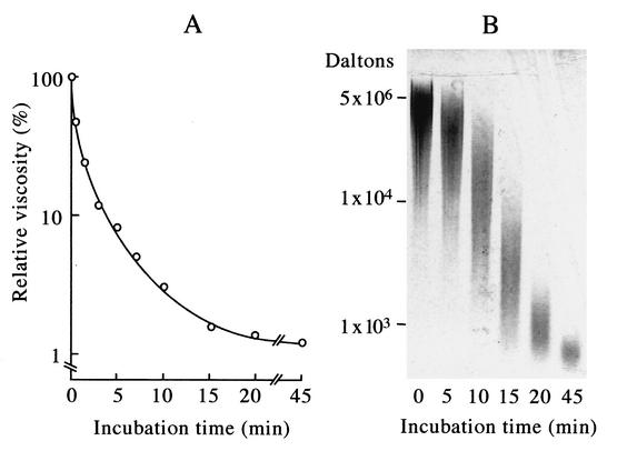
FIG. 1.
γ-PGA degradation activity in a B. subtilis NAFM5 culture infected with ΦNIT1 phage measured by viscometry (A) and by agarose gel electrophoresis (B). Reactions proceeded as described in Materials and Methods, and portions of mixtures were withdrawn after the indicated incubation periods. (A) Viscosity was measured using a falling-ball viscometer and is expressed as relative viscosity to the mixture at zero time. (B) Samples (10 μl) were resolved by 1.0% agarose gel electrophoresis, and degradation products were visualized by staining with methylene blue. Positions of intact γ-PGA (5 × 10⁶ Da) and of 104 Da and 103 Da γ-PGA (fractionated from partial hydrolysates of γ-PGA by PghP through gel filtration and high-performance liquid chromatography) (21) are indicated at left.

