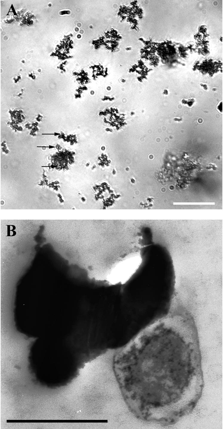
FIG. 4.

Light (A) and electron (B) microscopic images of FeOB (strain FO10, a member of the γ-Proteobacteria). Cells were grown anaerobically with ferrous carbonate (FeCO3) and nitrate (NO3−). Bars, 20 μm (A) and 0.5 μm (B). Irregularly twisted Fe oxide particles are apparent by light microscopy in this culture. By electron microscopy, cells can be seen closely associated with Fe particles, which appear to nucleate at the cell surface or within a capsule-like structure that surrounds cells. The close association between cells and mineral particles can be seen by light microscopy; cells appear as roughly circular open areas within mineral aggregates (arrows in panel A).
