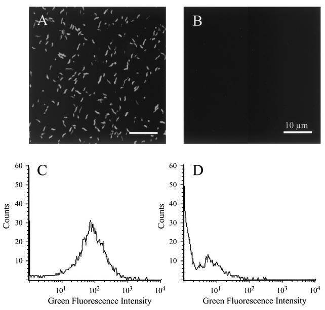
FIG. 2.
Green fluorescence of C. jejuni harboring pMEK91. C. jejuni was cultured on MHB agar plates, harvested in PBS, and visualized with a confocal microscope (A and B). In addition, bacterial samples were suspended in PBS and analyzed by flow cytometry (C and D). Data for the C. jejuni F38011 isolate expressing gfp are shown in panels A and C, whereas the data for the nontransformed C. jejuni F38011 wild-type isolate are shown in panels B and D.

