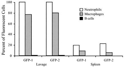
FIG. 3.
C. jejuni synthesizing GFP displays a host cell association in the peritoneal lavage and spleen. Four hours after intraperitoneal injection with either PBS or bacteria, mice were euthanized, and cell suspensions were prepared for flow cytometric analysis. The subset of cells expressing the appropriate surface markers was analyzed to determine the proportion of neutrophils (CD11b+ Gr-1+), macrophages (CD11b+ Gr-1−), and B lymphocytes (CD45R+ CD11b−) with green fluorescence above background. The data presented were collected from two mice injected with C. jejuni synthesizing GFP (GFP-1 and GFP-2).

