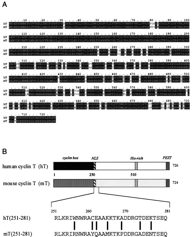
Figure 1.
Comparison of human and mouse cyclin T proteins. (A) Alignment of sequences from the human and mouse cyclin T proteins. Identical and similar amino acids are depicted on black and gray backgrounds, respectively. These two cyclins share 90% sequence identity. (B) Schematic representation of cyclin T proteins. Of note are duplicated cyclin boxes from positions 1 to 250, the putative nuclear localization signal (NLS) from positions 251 to 255, the histidine-rich region (His-rich) from positions 506 to 530, and the C-terminal PEST sequence from positions 709 to 726. The shading between these cyclins is slightly different for easier reference in Figs. 2 and 3. Sequences flanking cyclin boxes from positions 251 to 281 are presented below the schematic representation. Seven amino acids differ between the human and mouse cyclin T. Of these, the cysteine, lysine, alanine, and lysine at positions 261, 265, 269, and 277 in human cyclin T were changed individually to tyrosine, methionine, proline, and asparagine from mouse cyclin T, respectively (see below).

