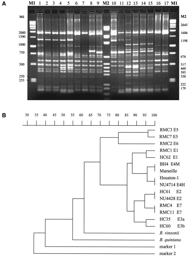
FIG. 4.
ERIC-PCR. (A) Representative gel electrophoresis of ERIC-PCR. Lanes 1 to 3, pattern E4H (strains ATCC 49882 [Houston-1] and NU4714); lanes 4 and 5, pattern E4M (strains BH4 and Marseille); lanes 6 and 7, pattern E2 (strains HC61 and NU4428); lanes 8 and 9, patterns E3a and -b, respectively (strains HC35 and HC60); lanes 10 and 11, pattern E1 (strains HC62 and RMC1); lanes 12 and 13, pattern E5 (strains RMC3 and RMC7); lanes 14 and 15, pattern E6 (strain RMC2 duplicate); lanes 16 and 17, pattern E7 (strains RMC11 and RMC4); lanes M1 and M2, molecular size markers. (B) Dendrogram. B. quintana and B. vinsonii patterns are quite distinct from all others (not shown in panel A) and are included in a merged reconstructed dendrogram. The dendrogram reflects the similarity of distribution of ERIC priming sites and is derived from gels (including that shown in panel A), all of which included B. henselae and the two molecular size markers and were analyzed identically.

