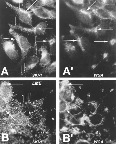
Figure 5.
hSKI-1 immunoreactivity in stably transfected HK293 cells. Black and white representation of the comparative double (red and green) fluorescence staining using an SKI-1 antiserum (directed against amino acids 634–651) (A and B) and fluorescein isothiocyanate-labeled WGA (A′ and B′) in control (A and A′) and leucine methyl ester (LME)-treated (B and B′) cells. Thin arrows emphasize the observed punctate staining, which is enhanced in the presence of LME. Large arrows point to the coincident staining of SKI-1 and WGA. [×900; bar (B′) = 10 μm.]

