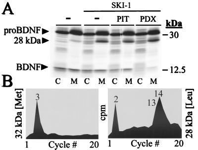
Figure 6.
Processing of proBDNF by SKI-1. (A) COS-7 cells were infected with vv:BDNF and either vv:WT (−) or vv:SKI-1 in the presence of either vv:PIT or vv:PDX. The cells were labeled metabolically with [35S]cysteine/[35S]methionine for 4 h, and the media (M) and cell lysates (C) were immunoprecipitated with a BDNF antiserum before SDS/PAGE analysis. The autoradiogram shows the migration positions of proBDNF (32 kDa), the 28-kDa BDNF produced by SKI-1, and the 14-kDa BDNF. (B) Microsequence analysis of the [35S]Met-labeled 32-kDa proBDNF (maximal scale, 1,000 cpm) and [3H]Leu-labeled 28-kDa BDNF (maximal scale, 250 cpm).

