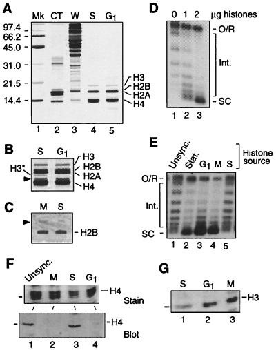
Figure 4.
Modification of histones H3 and/or H4 regulates replication-independent chromatin assembly. (A) Analysis of histones isolated from cdc mutants arrested in S phase (cdc17–1) and G1. Acid-extracted yeast histones (1 μg per lane) were resolved by SDS/PAGE and visualized by silver staining. The protein profile of calf thymus histones (CT) and whole cell extract (W, 20 μg) is shown for comparison. Note the faint staining of H3 in calf thymus and yeast histones. The migration of protein standards (Mk) in kDa is indicated on the left. (B) Detail of lanes 4 and 5 in (A) after prolonged staining. H3* is a presumed degradation fragment of H3. The arrowhead indicates an unidentified protein. (C) Immunostaining of H2B in histones isolated from M (cdc15) and S phase (cdc17–1) extracts. Proteins (20 μg) were resolved by triton-acid-urea gel electrophoresis and probed with anti-H2B antibody. The arrowhead indicates a cross-reacting protein also detected by the preimmune serum. (D) Add-back of histones purified from late log phase assembly extract stimulates assembly in test extract (50 μg). Reactions were performed at 30°C for 30 min. The migration of open circular/relaxed DNA (O/R), highly supercoiled species (SC), and intermediate topoisomers (Int.) is indicated. (E) Direct comparison of assembly capacity of purified histones from assembly extracts of unsynchronized log phase cells (Unsync.), stationary phase cells (Stat.), and cdc mutants arrested at specified points in the cell cycle. Reactions performed as in C. Posttranslational modifications of histones H3 and H4 in yeast chromatin assembly extract. (F) Histones (20 μg) isolated from M (cdc15–1), S (cdc17–1), and G1 (cdc28–1) extracts were resolved by SDS/PAGE and detected by Coomassie blue staining [Upper; only proteins similar to H4 (the middle band of the triplet) in size are shown] or characterized by Western blot analysis using an antibody raised against the tetra-acetylated isoform of the amino-terminal tail of H4 (Lower). (G) Western blot analysis as in F but with an antibody raised against the phospho-Ser 10 isoform of the amino-terminal tail of H3.

