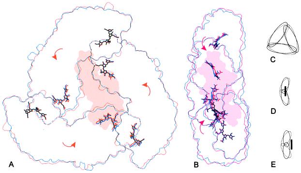
Figure 4.
Comparison of the molecular outlines of the symmetric, linker-free allophycocyanin (12) and the linker-containing complex (A and B) as well as an exaggerated schematic representation (C–E): The symmetric allophycocyanin (blue or dashed lines) is compared with molecule M (orange) in front view (A) and molecule N (red) in side view (B). On binding of the linker, the entire complex collapses toward the pseudo-threefold axis (C) and the α/β-ring becomes flattened (D and E) because of a rotation of the monomers as indicated by the arrows. In molecule M the linker inserts further into the trimer (D), resulting in a larger movement of the chromophores toward the pseudo-threefold axis, but with a slighter displacement of the chromophores to the right as compared with molecule N (E). These outlines were prepared by using an edge-detection filter and grasp surface representations of each molecule (50).

