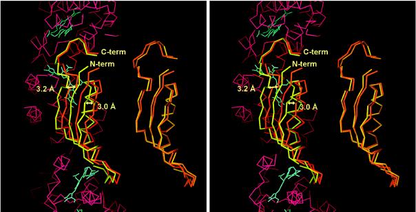
Figure 5.
Comparison of the structure of the linker polypeptides of the two molecules in the asymmetric unit. On the right the linker Cα-atoms are aligned directly by an rms procedure. On the left, the linkers are shown after rms alignment of the Cα-atoms of the two trimers in the asymmetric unit. The two linker molecules are very similar. However, the linker in molecule M (yellow) is shifted by about 3 Å to the left with respect to the molecule N linker (orange). Part of the Cα-trace, as well as the chromophores of molecule N, is shown as reference in red and blue, respectively. The figure was prepared in main (29).

