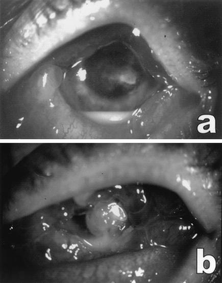
FIG. 1.
Left eye of patient. (a) On presentation. An infiltrate was present in the central part of the cornea and a hypopyon was visible in the anterior chamber. The conjunctiva was heavily injected. (b) Three days after presentation. Shown are increased corneal infiltrate and hypopyon in the anterior chamber, after treatment with amphotericin B and itraconazole eyedrops. Note the severe edema and hyperemia of the conjunctiva.

