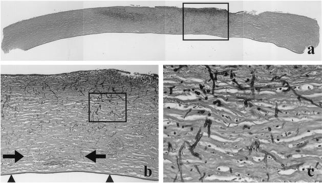
FIG. 2.
(a) Composition of a cross-section of the cornea button; the darker areas in the cornea stroma represent inflammatory infiltrate with necrosis and fungi. Note that the periphery of the cornea does not show this change. (b) Shown is a higher magnification of the area indicated by the rectangle in panel a. It illustrates that some fungi (arrows) and inflammatory cells reach close to Descemet's membrane (arrowheads); however, penetration of this membrane was not found. (c) Shown is the area indicated by the rectangle in panel b. The fungi appear as septated, branching hyphae with variable diameter. PAS staining is shown in all panels. Original magnifications, ×50 (a), ×100 (b), and ×400 (c).

