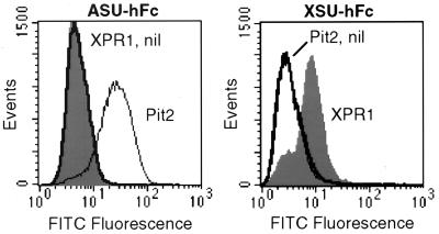
Figure 1.
SU-immunoadhesin binding to CHO cells expressing XPR1 or Pit2. Cells were transduced with L(XPR1)SN (XPR1, filled histograms), L(Pit2)SN (Pit2, thin lines), or the LXSN vector without an insert (nil, thick lines) and were selected in G418 to generate polyclonal populations of vector-expressing cells. The cells were detached by treatment with 1 mM EDTA, were washed with PBS, were incubated with ASU-hFc (Left) or XSU-hFc (Right) for 30 min at 37°C, were washed twice with PBS, were incubated with rabbit F(ab′)2 anti-human IgG conjugated to fluorescein isothiocyanate (Dako), and were washed before flow cytometric analysis. Dead and clumped cells were excluded by gating on forward and high-angle light scatter and exclusion of propidium iodide (1 μg/ml), and 50,000 gated events were analyzed per sample.

