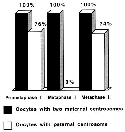
Figure 2.
Quantitation of maternal and paternal aster formation in Spisula embryos during meiosis I and II. Oocytes were fertilized, were fixed at various times, and were processed for immunofluorescence analysis. Over 100 embryos were analyzed for maternal and paternal aster content at each stage. Ten minutes after fertilization (prometaphase I), all embryos contained two maternal asters, and 76% contained one paternal aster associated with the sperm nucleus. Twenty minutes after fertilization (metaphase I), all embryos contained two maternal asters, but no paternal asters were identified in any of the embryos analyzed at this time point. Forty minutes after fertilization (metaphase II), all embryos contained two maternal asters, and 74% contained one paternal aster.

