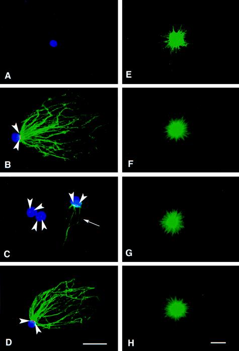
Figure 3.
In vitro assembly of paternal and maternal asters. Immunofluorescence analysis of isolated sperm heads (A–D) or isolated maternal centrosomes (E–H) incubated in buffer (A and E) or oocyte lysates (B–D and F–H) by using anti-γ-tubulin antibody (red), antitubulin antibody (green), and ethidium homodimer to stain chromatin (blue). (A) No γ-tubulin or Mts were found associated with sperm heads incubated in buffer. (B) Paternal centrosomes acquired both γ-tubulin staining (arrowheads) and the ability to nucleate Mts when incubated in 10-min lysate. (C) When incubated in 20-min lysate, the paternal centrosomes showed very weak γ-tubulin staining (arrowheads) and little or no Mt nucleation potential (arrow). (D) When treated with 40-min lysate, the paternal centrosomes nucleated Mts and displayed obvious γ-tubulin staining (arrowheads). (E–H) No obvious difference in the Mt nucleation potential of maternal centrosomes was observed as a function of buffer or lysate treatment. [Bars = 5 μm (D, for A–D, and H, for E–H).]

