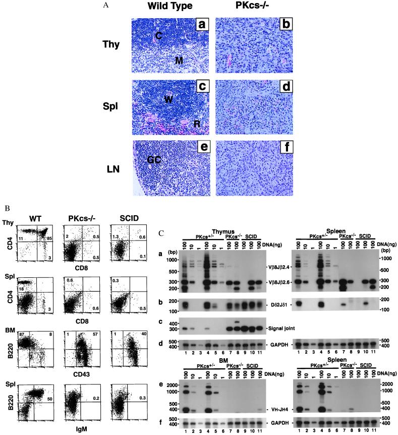
Figure 2.
Development of lymphocytes is blocked at early stages in DNA-PKcs−/− mice. (A) Histological analysis of thymus (Thy), spleen (Spl) and lymph node (LN) from wild-type and DNA-PKcs−/− mice (×200). Tissue sections were stained with hematoxylin and eosin. In tissue samples from DNA-PKcs-deficient mice, we observed effacement of normal histology and replacement by immature cells. The abbreviations are as follows: C, cortex; M, medulla; W, white pulp; R, red pulp; GC, germinal center. (B) Flow cytometric analysis of cells from the thymus (Thy), bone marrow (BM), and spleen (Spl) for the presence of precursor and mature T and B cells. Thymocytes and splenocytes were stained with fluorochrome-conjugated antibodies to CD4 and CD8; splenocytes and BM cells were stained with fluorochrome-conjugated antibodies to B220 and IgM or CD43. Profiles shown are representative results from a 4- to 5-week-old DNA-PKcs−/− mouse, its heterozygous littermate, and an age-matched CB-17 SCID mouse. (C) TCR and Ig gene rearrangement in DNA-PKcs−/− mice. (a) TCRβ rearrangement by PCR analysis. Thymus and spleen DNA were assayed for recombination of Vβ8-Jβ2.6. Both the quantity and the diversity of TCRβ rearrangement were reduced in DNA-PKcs−/− and SCID mice. (b) Coding joint of TCRδ rearrangement. Thymus and spleen DNA were assayed for recombination of Dδ2-Jδ1. (c) Signal joint of TCRδ rearrangement. Thymus DNA was assayed for Dδ2-Jδ1 circular signal joint products. There is more amplified signal for both DNA-PKcs−/− and SCID mice than for heterozygous control mice. (e) Ig heavy-chain rearrangement by PCR analysis. BM and spleen DNA were used for recombination of VH7183-JH4. Rearrangement in DNA-PKcs−/− and SCID is severely reduced in both BM and spleen. (d) and (f) Control GAPDH amplification from thymus, spleen, and BM DNA. DNA (100, 10, or 1 ng) from thymus, spleen, and BM or from a 5-week-old DNA-PKcs+/− mouse (lanes 1–3), of a 9-week-old DNA-PKcs+/− mouse (lane 4–6), and 100 ng DNA of three individual DNA-PKcs−/− mice (lanes 7–9) and three individual SCID mice (lanes 10–12). DNA-PKcs−/− and SCID mice analyzed were also between 4 and 9 weeks of age.

