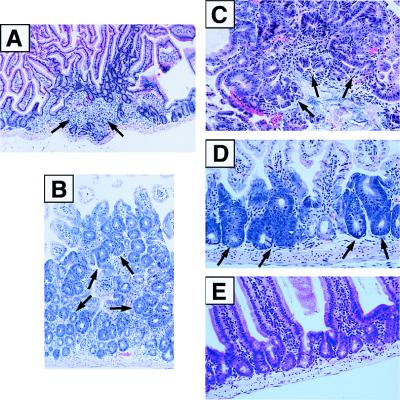
Figure 4.
Preneoplastic lesions in DNA-PKcs−/− mice. Intestinal tissue samples from 6-week- to 6-month-old DNA-PKcs−/− mice were sectioned, stained with hematoxylin and eosin, and photographed. (A) Section of intestinal tissue showing inflammation and mild epithelial hyperplasia (×100). (B) Photomicrograph of colonic mucosa showing crypt hyperplasia with mild to moderate dysplasia (×200). (C) Adenomatous polyp of the colon showing areas of severe dysplasia (×400). (D) Aberrant crypt foci along the intestinal mucosa showing severe dysplasia (×400). (E) Section of intestinal tissue from a wild-type mouse showing normal morphology (×250).

