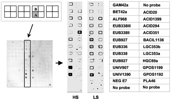
FIG. 1.
DNA microarray images hybridized with rRNAs extracted from sediment samples collected at a high-salinity (HS) site and a low-salinity (LS) site, and position of the probes on the microarray. The microarrays were hybridized and washed at 20°C. Each probe was spotted in duplicate on the microarray. DNA microarray images of quadrant A are shown since quadrant B yielded virtually identical results. Probe names are listed in Table 1.

