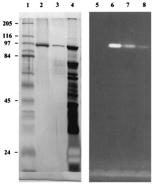
FIG. 4.
Purification of β-mannosidase (ManB) as demonstrated by SDS-PAGE. A silver-stained gel is shown on the left. Lanes: 1, broad-range marker (Sigma); 2, purified ManB eluted from Superose-12 column; 3, partially purified mannosidase eluted from Mono-Q column; 4, crude enzyme preparation. Zymograms demonstrating the activity of the same enzyme fractions using MUβMan as substrate are on the right. Lanes: 5, broad-range marker (Sigma); 6, purified ManB eluted from Superose-12 column; 7, partially purified mannosidase eluted from Mono-Q column; 8, crude enzyme preparation. Molecular masses are shown at right (in kilodaltons)

