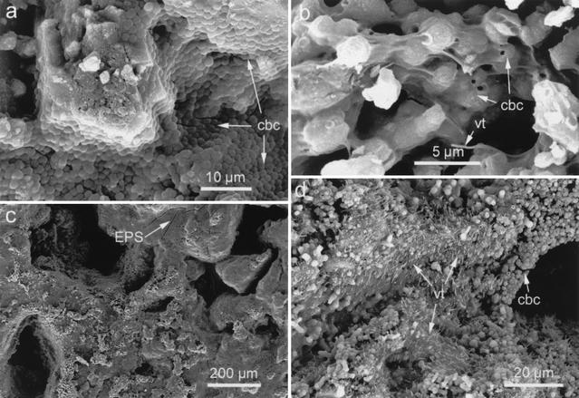
FIG. 6.
SEM photomicrographs of large samples cultured under nonshaking conditions. (a) Calcified bacterial cells (cbc) covering the pore walls of samples cultivated in the M-3 medium (after 30 days). (b) Detail of calcified bacteria linked by needle-like vaterite (vt) crystals in samples cultured in the M-3P medium (after 10 days). (c) Sample cultured in the M-3P medium (after 30 days) showing incipient development of an EPS film (the arrow indicates cracks in the film; note that no pore plugging occurred). (d) Massive needle-like vaterite crystals blanketing stone pores following 30 days of culture in the M-3P medium. Calcified bacterial cells are also observed covering the pore walls.

