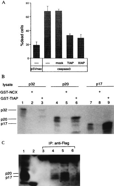
Figure 2.
Inhibition of apoptosis induced by overexpression of caspase and association with processed form of caspase 3. (A) Rat-1 cells were cotransfected with plasmids expressing caspase 3 and a plasmid expressing Tiap or Xiap and cell death was measured. (B) [35S]Methionine-labeled in vitro-translated p17, p20, or p32 of caspase 3 was mixed with GST-TIAP or GST-NCX, as indicated, followed by precipitation with glutathione-agarose beads and analysis by SDS/PAGE. In vitro-translated proteins were directly applied to lane 1, 4, or 7. (C) Jurkat cells stably transfected with Flag-tagged TIAP (lane 4), XIAP (lane 5), or control vector (pCR2FL) (lane 6) were stimulated with anti-Fas antibody and immunoprecipitated with anti-Flag antibody. Immunoprecipitates were analyzed by immunoblot with anti-caspase 3 antibody. Lanes: 1, lysate from Jurkat control transfectant stimulated with anti-Fas antibody; 2, marker; 3, Jurkat parent cells.

