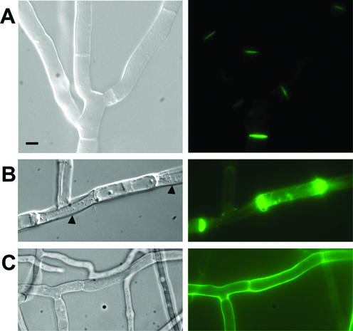
FIG. 1.
Localization of the IDI-1-GFP fusion protein. (A) Strain expressing the IDI-1-GFP fusion protein and grown on rich medium. (B) Incompatible het-R het-V strain expressing the IDI-1-GFP fusion protein and transferred for 4 h at 26°C (incompatibility conditions). The black arrowheads mark lysed cells surrounding a surviving cell. (C) Strain expressing the IDI-1-GFP fusion protein grown on nitrogen-deprived medium. For each panel, the Nomarski view is given on the left and the GFP fluorescence view is given on the right. Note that image acquisition time was 10 times longer for panel A than for panels B and C. The scale bar represents 2 μm.

