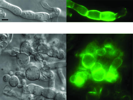
FIG. 6.
Rapamycin induces morphological defects and expression of the IDI-1-GFP fusion protein. A strain expressing the IDI-1-GFP fusion protein was grown on medium containing rapamycin (500 ng · ml−1). The Nomarski view is given on the left, and the GFP fluorescence view is given on the right. The scale bar represents 2 μm. Note the abnormal cellular morphology induced by rapamycin. Image acquisition time was 50 times shorter than that for Fig. 1A.

