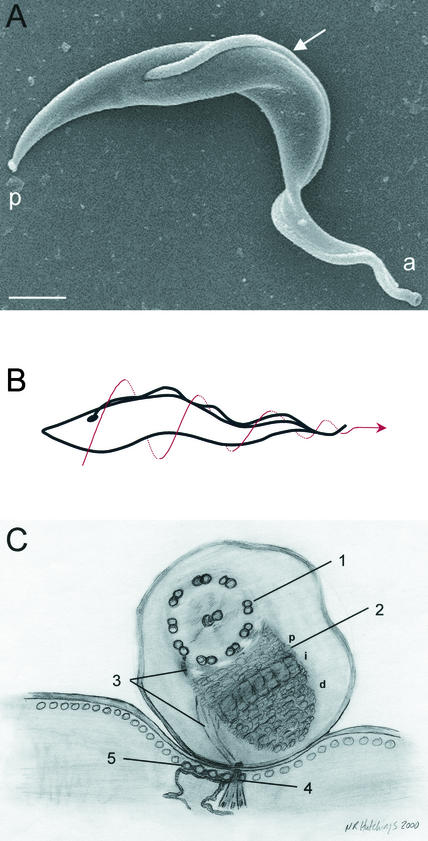
FIG. 1.
T. brucei cell structure. (A) Scanning electron micrograph of T. brucei. The flagellum (arrow) is attached to the cell body along its length. The flagellum emerges from the posterior (p) end of the cell and charts a left-handed helical path as it extends toward the anterior end of the cell (a). Bar, 1 μm. (B) Cartoon depicting the auger-like movement of the trypanosome cell. The dotted arrow indicates cell motion. (C) Schematic illustration of a transverse section through the FAZ, as viewed from the posterior end of the cell. The flagellum (top) contains a canonical 9 + 2 axoneme (1) and the PFR (2). A network of thin filaments (3) connects the axoneme and PFR to the cytoplasmic FAZ filament (4). Together with a specialized quartet of reticulum-associated microtubules (5), these filaments comprise the FAZ, which extends from the flagellar pocket to the anterior end of the cell. See the text for further details. The proximal (p), intermediate (i), and distal (d) regions of the PFR are indicated. (Reprinted from references 28 and 33 with permission of the publishers.)

