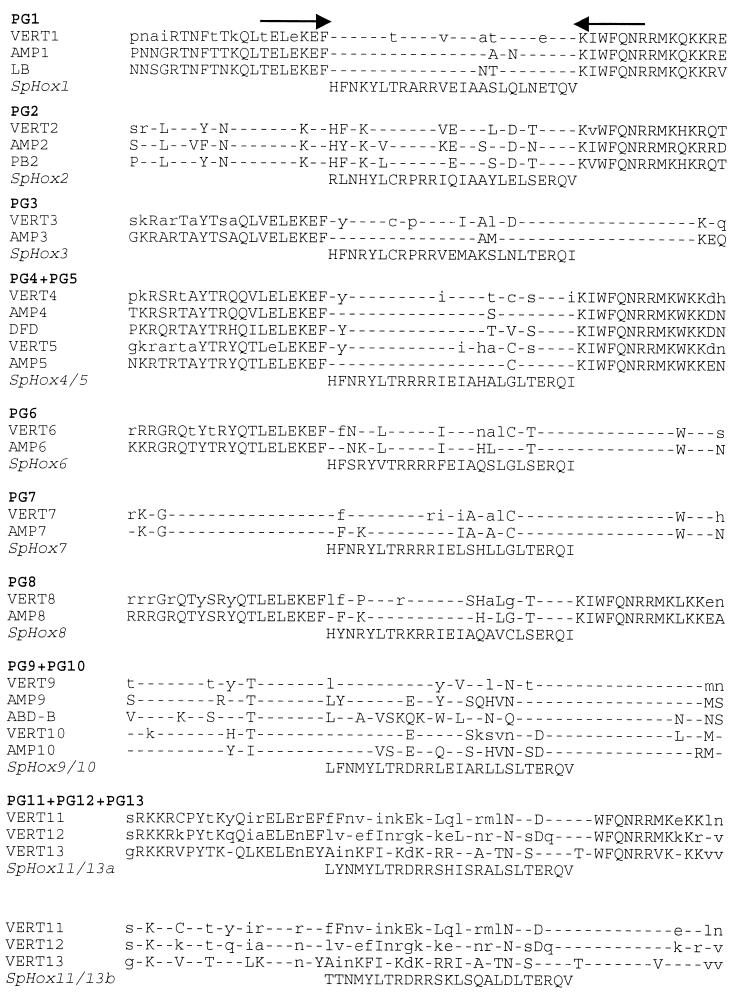
Figure 2.
Alignment of vertebrate, amphioxus, Drosophila, and S. purpuratus homeodomain sequences. The S. purpuratus homeodomain sequences are shown flanked by arrows representing the positions of the PCR fragments used in our screens (18). Homeodomain sequences for some genes were obtained by other methods or were from a combination of sources, e.g., clones isolated by genomic walking or cDNA clones, as described in the text. In vertebrate homeodomain consensus sequences (VERT), uppercase letters indicate a residue conserved in all known vertebrate sequences of that paralog group, e.g., all mouse and human PG1 genes (24). Lowercase letters indicate a residue found in the majority but not all vertebrate sequences of each paralog group, i.e., comparing the multiple vertebrate sequences available for each Paralog Group (there is only a single amphioxus gene from each Paralog Group). Dashes indicate amino acid identity at that position between the S. purpuratus genes and all vertebrate genes as well as Drosophila and amphioxus genes of that paralog group. Amphioxus sequences [AMP, from Branchiostoma (3)] are shown below the vertebrate consensus sequences. Drosophila sequences included in the comparison are Labial (LB), Proboscipedia (PB), Deformed (DFD), and Abdominal B (ABD-B). Sequences are compiled from ref. 25.

